Millions of people are diagnosed with colorectal cancer yearly, and over 25,000 are here in Canada. Part of diagnosing certain types of cancers require pathologists to search for lymph nodes in resected cancer specimens by hand, which makes the process labour, time, and resource intensive. Crucially, given the limitations of human touch, smaller lymph nodes may be missed.
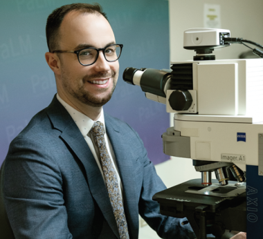
Matthew Cecchini, a medical doctor and assistant professor in pathology and laboratory medicine is part of a team of researchers that have developed a novel technology dubbed the Lymphonator. “I work as an anatomical pathologist specializing in pulmonary, head and neck and molecular pathology, and I also participate on the gastrointestinal and pediatric pathology teams. Additionally, I am interested in developing and utilizing digital pathology tools to enhance clinical practice,” said Cecchini.
The Lymphonator is a bench-top robotic scanning device that guides a hospital’s pathology team in efficient and reliable lymph node identification in surgically removed colorectal cancer tissues. This helps eliminate inefficiencies in the pathology workflow, optimize cancer care, and reduce healthcare costs.
The device has an ultrasound imaging tool that scans the surgically removed cancer specimen and identifies the lymph nodes for extraction. As the process has been automated, pathology staff can configure the device to perform the scan independently, freeing them up to perform their other clinical duties simultaneously. The device is also susceptible, detecting even the most minor lymph nodes resulting in higher accuracy in forming treatment decisions and lowering patient complications and mortalities.
The invention has been licensed by Tenomix Inc., a startup founded by a group of former Western Medical Innovation Fellows. At Tenomix, their vision is to be at the forefront of pathology innovation, ensuring pathology staff are equipped with the right tools that help provide the best patient care.
Read more


